Topic 1: The Eukaryotic Cell

A eukaryotic cell is defined as a cell with a nucleus and membrane-bound organelles.  Animal and plant cells are eukaryotic, but a slightly different from each other.  This is their appearance:

Generalized animal cell


Image from:  http://www.bio.miami.edu

Generalized plant cell

  Image from:  http://www.uvm.edu


Structure and function of the cellular organelles:

1.  Cytoplasm:  the matrix in which all cell organelles and inclusions (insoluble waste or storage products) are found.  Made up of the cytosol and cytoskeleton.
-cytosol:  90% water, site of many biochemical reactions
-cytoskeleton:  made up of microtubules and microfilaments (string-like proteins).  Important in supporting the cell, giving its shape and some cell movements (like extension and contraction of pseudopods in amoeba!), as well as moving organelles within the cell and cell division.  The microtubules form parts of the cilia and flagella.


2.  Nucleus:  control center of most cellular activities.  It is surrounded by the nuclear envelope, which contains pores though which materials (proteins and RNA) can pass.  The nucleoplasm is found within the envelope, it is similar to the cytoplasm.  The nucleolus is the dark patch in the nucleus where ribosomal RNA is formed.  Chromatin (DNA and associated proteins) is found around the nucleolus.


3.  Endoplasmic reticulum (ER):  It is a series of interconnected, flattened sacs, tubes and channels formed by the phospholipid bilayer.  There are 2 types:  rough ER and smooth ER.  Rough ER appears bumpy because it is covered with ribosomes.  Its function is to isolate, store and transport proteins produced by the ribosomes.  The smooth ER produces and stores lipids.

4.  Golgi apparatus:  also made of phospholipid bilayers (or membranes), it functions to modify, sort and package macromolecules (like proteins and lipids)  for cell secretion (exocytosis) or use within the cell.  Imagine it is like a post office; it packages and labels items and sends them to different parts of the cell.


5.  Ribosomes:  make proteins from amino acids.  They can be found attached to the ER or floating freely in the cytoplasm.  They are made of rRNA and protein.

In the following picture, the connection between the various cell parts can be observed.  Information about how to build a protein comes from the nucleus as mRNA.  It attaches to the ribosome, which "reads" the mRNA and builds a protein from amino acids.  The protein is released into the rough ER, then it is packaged in a vesicle (this is a little membrane-bound bubble) and transported to the Golgi apparatus, where it may be modified, packaged into a new vesicle and then transported to the cell surface and released (exocytosis).

Image from:  http://scienceblogs.com/transcript/upload/2006/07/ergolgi.jpg

6.  Mitochondria:  small membrane-bound organelles in the cytoplasm.  They are considered the "powerhouse" of the cell as this is where the reactions occur that create ATP (cellular respiration) that provide the cell with energy.

 Image from:  http://imagineannie.files.wordpress.com/2009/11/mitochondria1.jpg


 Image from:  http://math.etsu.edu/symbiosis/mitochondria.jpg


7.  Chloroplasts:  Found only in plants and plant-like protists.  They are the site of photosynthesis and contain the green pigment, chlorophyll (along with other, less visible pigments!)

 Image from:  http://www.scsc.k12.in.us/SMS/Teachers/Martin/chloroplast.jpg

  Image from:  http://botit.botany.wisc.edu/images/130/Photosynthesis/Chloroplast_EN.gif


8. Lysosomes and peroxisomes:  Special vacuoles, which are designed to digest things (such as worn-out or excess cell parts, food particles, invading viruses or bacteria, etc) with digestive enzymes.  They contain different digestive enzymes, therefore digest different things.  Lysosomes are formed from the Golgi apparatus, whereas peroxisomes are formed from the ER.

9.  Cell wall:  It is found in plant, algal and fungal cells.  It is completely permeable, but it contains cellulose (plants and algae) or chitin (fungus), which protects and supports the cell.


10.  Cell membrane:  It is a phospholipid bilayer.  It is both elastic and rigid and helps give the cell its shape.  It is selectively permeable, thus helps control transport of substances and homeostasis.  It is permeable to gases and water, but many substances can only pass through it with the help of proteins, such as channels, protein pumps and receptor proteins.  

 Image from:  http://macro.magnet.sfu.edu/cells/plasmamembrane/images/plasmamembranefigure1.jpg


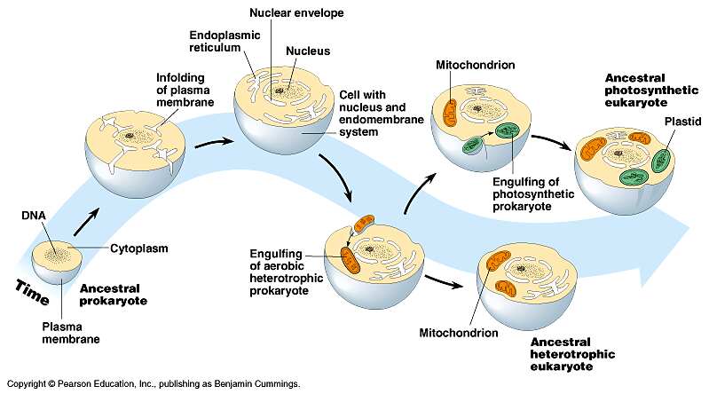
The Endosymbiotic Theory
-  proposed by Lynn Margulis at the end of the 1960s, it was initially ridiculed, but is now well-accepted.
-   suggests that certain prokaryotic cells engulfed (endocytosed) other cells, but instead of being digested, these cells continued to live and each provided the other with some benefits (symbiosis).
-  mitchondria:  an anaerobic bacteria engulfed a smaller aerobic bacteria.  Mutual advantages?  The aerobic bacteria provided the larger, anaerobic bacteria with the ability to survive in areas with oxygen, while the larger bacteria ingested food and provided protection to the smaller aerobic one.
-  chloroplast:  a photosynthetic bacteria was engulfed by a larger anerobic bacteria.  Advantages?  I am sure you can figure these out for yourselves!!
-  evidence:  These organelles are surrounded by a double lipid bilayer (their own, plus the one of the cell that engulfed them!).  These organelles also have their own DNA and divide independently of the cells that they are found in.

 Image from:  http://www.origin-of-mitochondria.net/?attachment_id=120

Topic 2: Transport across the membranes


The plasma membrane of a cell has many functions:
-separate internal and external environment
-protect the cell
-a place for recognition sites
-controls transport processes
As such, it is rigid, yet elastic and selectively permeable (this means it lets some things cross through it, but others cannot)

Structure of the plasma membrane
- 7-9nm thick (under an electron microscope it only looks like 2 lines)
- organic solvents (eg. alcohol, ether, chloroform) can cross it, since it is made of lipids
- in 1959, the unit membrane theory was created, which stated that all membranes (surface and inner) share a basic structure.  This is true.  It also stated that it was 2 protein layers (for elasticity) sandwiching a phospholipid bilayer (for strength).  This was incorrect.

- 13 years later (1972) a technique called "freeze-fracture" was used to visualize the membrane and it showed that the unit membrane theory was WRONG!  So a new theory called the fluid mosaic model was created (and so far still seems right!).

It states that there is:
-a phospholipid bilayer in which the phospholipids can move (hence the fluid part of the name)
-cholesterol (steroid lipid) is found interspersed in the bilayer.  Cholesterol is more rigid than phospholipids, so it provides stability.
-globular membrane proteins are embedded in the membrane, but they don't form a continuous layer.  Different proteins have different functions, some are transport molecules (they create channels through the bilayer, or they carry certain substances across the bilayer), some are enzymes, some are receptor molecules.  Proteins can also move sideways in the membrane.  Proteins provide the mosaic part of the fluid-mosaic model.
-carbohydrates are found on the outside only.  They form receptor sites and are attached to proteins or lipids (glycoprotein, glycolipid).

 Image from http://www.biology.arizona.edu/cell_bio/problem_sets/membranes/graphics/fluid_%20mosaid_model.jpg


 Transport Processes
- homeostasis maintains constant conditions in a living organism.  Membrane control is essential for this.

Passive transport
This is movement across the membrane without any energy input.  There are 3 forms:  diffusion, facilitated diffusion and osmosis

Diffusion:
- particles are in constant motion (Brownian or heat-kinetic motion)
-the overall effect of this random motion over time is diffusion (or the movement of substances from an area of high concentration to an area of lower concentration, thus resulting in equal concentration in both areas)
-for this to occur across the membrane, the membrane must be permeable to the substances (apolar particles and small molecules like H2O, O2 and CO2)

Facilitated diffusion:
-a transport protein helps the molecule to pass through the membrane - these proteins usually form channels through the membrane (some may have gates to limit diffusion) and some are carrier proteins
-changed ions or polar particles can only move across the membrane with the help of a protein.

Osmosis:
-diffusion of H2O molecules (a special kind of diffusion)
We can observe osmosis by placing cells into different solutions:
An isotonic solution has the same concentration as a cell, so no visible changes occur (this is called dynamic equilibrium)
A hypotonic solution is less concentrated than the cell, so in this case the cell swells and bursts (or in the case of a plant cell, the cell walls prevent bursting, so there is an increase in turgor pressure)
A hypertonic solution is more concentrated than the cell, so the cells shrivels up (in plants this also happens and the cell membrane pulls away from the cell wall, this is called plasmolysis).
 Image from http://access.mmhs.ca/docs/Science/MMHS%20Web%20Folder/Kamla/Image130.gif


Active Transport
- transport of a substance against the concentration gradient
-requires energy
-must use transport proteins and ATP
-a substance binds the transport protein and energy is released from ATP to allow the protein to change shape or move and release the substance on the other side of the membrane.
Example:  Na+/K+ pump - this is especially important in nerve and muscle cells
Image from http://bio100.nicerweb.com/doc/classbio1151/Locked/medi/ch07/07_16SodiumPotassiumPump.jpg


Endocytosis and Exocytosis
- this occurs when large particles (even whole cells) move across the membrane.
Endocytosis occurs when something is moved into the cell.  The membrane engulfs and encloses the particle - this forms a vacuole or vesicle.
- 3 types:
phagocytosis:  engulfing a very large, solid particle
pinocytosis:  engulfing liquid droplets or small particles.
receptor-mediated endocytosis:  engulfing specific substances which bind to receptors found on the surface of the cell membrane.
Image from http://cellbiology.med.unsw.edu.au/units/images/endocytosis_types.png

Exocytosis occurs when something moves out of the cell.
-vesicles fuse with the membrane and dump out their contents
-used to expel wastes or secrete substances, such as hormones
 Image from http://jpke.nemc.edu.en/cb/kejian/3/lecture3.files/slide0001_image004.gif

Topic 3: Inner working of the cell: Water

In learning about the eukaryotic cell, proteins, carbohydrates, lipids and nucleic acids were mentioned.  What are these?  Well, the following topics will clarify that.


Only 25 of the 90 natural elements (periodic table) are essential for life.  We call these biogenic elements.
They can be divided into two groups:  macroelements (need lots) and microelements (needed in small quantities)

Macroelements
O - O2, H2O
C - CO2, organic compounds (lipids, carbohydrates, proteins, nucleic acids, vitamins)
H - H2O, CH4
N - amino acids, proteins, nucleic acids

H2O
-the most important inorganic compound
70-95% of most organisms (98% in jellyfish, about 70% in humans)
Image from www.brooklyn.cuny.edu/bc/ahp/SDgraphics/PSgraphics/SD.PS.LG.Water.html
- from the molecular structure you can see that water is a dipolar molecule with electron sharing
-hydrogen bond:  attraction of opposite charges, weak bond, H end attracts O end of another H2O or an anion
-very important in holding large molecules together, eg. proteins, DNA, etc

Properties:
1. Excellent solvent - H2O attracts ions and surrounds them, split ionic bond (dissociation) and prevents reassociation by creating hydrate shells (vizburok) around them, therefore they dissolve. eg. NaCl.  It also works for polar molecular substances eg. sucrose.  H2O can't dissolve apolar substances
2.Thermal properties: High thermal capacity (fajhő) - this is the amount of heat needed to raise the temperature of 1g by 1 degree Celsius.  Water has a high boiling point (liquid at ambient temperature and pressure)
3.  Surface tension (force that causes surface to contract to occupy the least possible area): Highest of any liquid except mercury, therefore water is pulled into spherical shapes and it creates a surface film, which supports insects, creates a habitat and causes capillarity (cohesion and adhesion) in plants' xylem.
4. Freezing properties: At temperatures LESS than 4C,  volume is high and density is low, therefore ice floats - this protects organisms that live in water.  At 4C water is densest and collects at the bottom.  Ice is a thermoinsulator.


Topic 4: Inner workings of the cell: Organic compounds

Organic compounds
-carbon based molecules, also called biomolecules

The role of carbon
Carbon has 4 electrons in its outer valence shell, therefore it can form very stable compounds by covalent bonding (a very strong type of bond).
The compounds formed can range from simple molecules to very large macromolecules.  They can be straight chains, branched chains or even rings, with single, double or triple bonds, therefore very diverse C skeletons.

 Image from www.contexo.info/DNA_Basics/Cell_Chemistry.htm
 Isomers increase diversity.  Isomers are compounds with the same atoms (chemical formula), but different 3D structures eg. glucose and fructose.


Image from faculty.clintoncc.suny.edu/faculty/michael.gregory/files/bio 101/bio 101 lectures/biochemistry/biochemi.htm

How large compounds are formed 
Monomers are smaller organic compounds which form the basis for the larger compounds.  They are often bound together in repetitive chains, therefore they are called the subunits of the larger compounds.
The larger compounds are called polymers.

Starch and glycogen are polymers made of many glucose monomers.

Polymers are formed by condensation reactions.

Hydrolysis breaks down polymers into monomers.

Images from www.bio.miami.edu


Topic 5: Inner workings of the cell: Carbohydrates (Szénhidrátok)

 Carbohydrates
-most common organic compounds (biomolecules)
-are energy sources for cells - broken down by cellular respiration (needs O2)-17kJ/g, half as much as lipids, but breakdown is much faster
-C,H,O
-can be mono-, di- and polyssacharides

a.  Monosaccharides
-sweet, H2O soluble, crystalizable
-general formula:  (CH2O)n
Triose sugars (n=3)
-these are all intermediate products (köztes termékek) in biochemical reactions
-most common: glyceraldehyde (oxidation product of glycerol)
Pentose sugars (n=5)
-biologically important: parts of nucleotides and nucleic acids, most common are deoxyribose and ribose


Image from http://www.layevangelism.com/bastxbk/images/deoxyribose.jpg

Hexose sugars (n=6)
-most common monosaccharides, eg. alpha-glucose, beta-glucose (szölőcukor), fructose (gyümölcscukor) and galactose are all isomers of C6H12O6 and form the monomers of most di- and polysaccharides
Image from http://www.nzetc.org/etexts/Bio14Tuat01/Bio14Tuat01_036a%28h280%29.jpg

I
Image from http://www.eastchester.k12.ny.us/schools/hs/teachers/fishman/images/fructose_000.gif


Galactose
Image from https://teach.lanecc.edu/naylore/225Lectures/04A/StructureImages/2galactoseThm.jpg


b.  Disaccharides
-2 monosaccharides combine by condensation to form a disaccharide
Examples:
glucose + fructose = sucrose (table sugar) - remember this is the main form in which carbohydrates are transported in plants!
I
Image from http://www.pharmas.co.uk/blog/wp-content/uploads/2009/04/glucose-fructose-and-sucrose.jpg

2 alpha glucoses = maltose - common in germinating seeds, result of hydrolysis (break down) of starch
I
Image from http://www.eastchester.k12.ny.us/schools/hs/teachers/fishman/images/maltose.gif
 
glucose + galactose = lactose (tejcukor) - found in milk
I
Image from http://dspace.dial.pipex.com/town/park/gfm11/nomilkgif/LACTOSE.JPG


c.  Polysaccharides
- largest carbohydrates
- long chains of monsaccharides (hexose sugars)
- can be folded
- general formula: (C6H10O5)n
- broken down into monomers (by hydrolysis) if needed for energy
- insoluble in H2O because of size
- not sweet
- cannot crystalize

TYPES of polysaccharides:
i.  Storage polysaccharides
-insoluble, therefore they won't diffuse out of cells and won't affect osmosis

Examples:
STARCH: found in plants - made of 2 types of chains
-amylose: unbranched alpha-glucose, stains blue with iodine
I
Image from http://www.cheng.cam.ac.uk/research/groups/polymer/RMP/nitin/Amylose1.jpg
-amylopectin:  alpha-glucose chain with side branches, stains red-purple with iodine
I
Image from http://www.vivo.colostate.edu/hbooks/pathphys/digestion/basics/amylopectin.gif
-the chains fold and pack together into starch grains (keményitőszemcse) - 20% amylose, 79% amylopectin, 1% fatty acids and phosphates

 GLYCOGEN: found in animals and fungi
- alpha-glucose is arranged like amylopectin (branched), but the chains are shorter and more branched.
-glycogen is found particularly in muscle and liver cells.
-human glycogen stores provide enough energy for a few hours.

ii.  Structural polysaccharides
Examples:
CELLULOSE
- makes up 50% of the plant cell wall
- each molecule is a chain of approximately 10 000 beta-glucose, unbranched
- OH-groups form H-bonds with neighbouring chains to create a lattice
- about 2000 chains mass together to form microfibrils, which are visible under an electron microscope

Image from http://www.botany.utexas.edu/facstaff/facpages/mbrown/newstat/stat38a.jpg
- the lattice is linked together by hemicellulose, which is a short polysaccharide
-cellulose is indigestible to animals (we consider it the "fiber" in our diet!)
-used to produce cotton, rayon, cellophane, celluloid and paper

CHITIN
- found in arthropod exoskeletons and fungal cell walls
- long chains of beta-glucose, but on each monomer the OH-group is substituted by a nitrogenous group (NHCOCH3)


Topic 6: Inner workings of the cell: Proteins

Proteins are large molecules.

They are not really soluble, rather they form colloids
- colloids are about 500nm, which is larger than particles in solution, but smaller than particles in suspension
- colloids exist in a "sol-gel state", whereby sometimes they appear to be liquid and at other times they are jelly-like (much of the material in cytoplasm is colloid)

Proteins contain: C, H, O and N, sometimes S and P

There are an almost limitless number of proteins, which vary between species and are often species-specific (fajlagosok).  They determine the characteristics of a species.

Types of Proteins
a.  Structural proteins - these form the organism. eg. hair, nails, feathers, etc.
b.  Physiological proteins - these carry out functions, examples include:
-enzymes (biocatalysts)
-carrier molecules (szállítómolekulák)
- pigments (eg. various colour molecules in skin and eyes, haemoglobin in red blood cells)
- hormones (chemical messengers)
- contractile material in muscles
- antibodies (disease protection)
** Proteins are rarely stored (only in seeds and eggs).  Proteins are only broken down for energy if a living organism is starving.

PROTEIN STRUCTURE
- a protein is a polymer.  Its monomers are called amino acids.


Image from http://api.ning.com/files/xO6ybWgUbfFlk7GUXm9d8dfR--U-fUdPOJEtDzVGgDY_/aminoacidstruc.jpg
- some amino acids are basic, others are neutral - this depends on the variable group
- some amino acids are polar and others are apolar - this depends on the variable group
-amino acids are soluble in water, where they form dipolar ions (zwitterion = ikerion), this means they have BOTH acid-base properties, so they have good buffering capacity.


Synthesis of polypeptides
- amino acids attach to each other by condensation to form covalent peptide bonds
2 amino acids condense to form a dipeptide, 3 form a tripeptide and many joined together form a polypeptide.
Képtalálat a következőre: „formation of dipeptide image”
Formation of a dipeptide
Image from:http://www.tutorvista.com/content/chemistry/chemistry-iv/biomolecules/chemical-properties--amino-acids.php

- if more than 100 amino acids attach together it is considered a protein
- polypeptides (and proteins) are broken down by hydrolysis
*both condensation and hydrolysis require enzymes to occur.

Structure
Primary structure: this is the number and sequence of the amino acids.
*Insulin was the first protein to have its primary structure determined by a researcher named Fred Sanger
Secondary structure: This type of structure is created by H-bonds forming between amino acid monomers
Alpha helix (eg. keratin - a major component of hair and skin)


Image from http://www.bio.miami.edu/~cmallery/150/protein/alpha-helix.jpg
Beta-pleated sheet (eg. silk protein)


Image from http://student.ccbcmd.edu/courses/bio141/lecguide/unit3/viruses/images/betasheet.jpg
-both structures can be found in a single protein.

Tertiary structure:  This is the secondary structure folded in 3-dimensional space.
-usually forms globular shapes
-bonded by S-bridges (requires the amino acid cysteine), ionic bonds, H-bonds and van der Waals forces


Image from http://lectures.molgen.mpg.de/ProteinStructure/Levels/tertiary.gif

Quaternary structure:  A protein has quaternary structure if it is formed of 2 or more subunits (polypeptides).  They are held together by various forces including hydrophobic interactions, H-bonds and ionic bonds.
eg. Haemoglobin

Image from http://www.theironfiles.co.uk/images/Haemoglobin_Structure.jpg

Proteins can further be catagorized as simple or complex.  A simple protein contains only amino acids, complex proteins often include other elements, such as the iron containing haeme molecule found in haemoglobin (above).

Protein Stability and Denaturation
A protein will be stable (maintain its shape and function) if the environment it is in is appropriate.  The most common environmental factors that will cause a protein to denature (lose its shape and/or function) are temperature and pH levels.  Some proteins have a wide range of tolerance (can function at 4C and at 40C), while others have a very narrow range.  This is a protein-specific characteristic.  An example of protein denaturation is when we cook an egg.  The white of the egg is almost entirely made of the protein albumin.  At room temperature it is a clear liquid.  If we increase the temperature, the protein starts to denature (lose its shape and therefore function too) and it become solid and white.  Denaturation occurs because the bonds between the amino acids are broken.
Sometimes denaturation is permanent (like cooking an egg), other times it can be reversible.

Topic 7: Inner workings of the cell: Lipids

Lipids are insoluble in water because they are nonpolar.
BUT, they do dissolve in apolar solvents, like alcohols, ethers, etc.

Lipids include:
Fats and oils
Phospholipids
Streroids
Carotenoids
Waxes

A. Neutral fats and oils
- these have high energy content
-structure:  made of a backbone with adjoining fatty acid chains
the backbone is an alcohol, commonly glycerol,


Image from lhs2.lps.org/staff/sputnam/Biology/U2Biochemistry/lipid_lab.htm

Fatty acids:  there is considerable variation in fatty acids, but basically they are hydrocarbon chains.  If all the bonds are single, then it is saturated, because it contains the maximum possible H's.  It will form a fat (solid).
The general formula is CH3(CH2)nCOOH.  
CH3 is the methyl group.
(CH2)n is the variable chain, for example if n=16, then it is a stearic acid chain (common in adipose tissue - zsírszövet).
COOH is the carboxyl group.
If the chain contains double bonds, then it is unsaturated.  It will form an oil (liquid).


Cells produce fats and oils by condensing (see lecture 1) a glycerol and 3 fatty acids.  These molecules are also called triglycerides.


Image from http://www.biology-books.com/Standard/ch2/16.jpg

The fatty acid part forms long apolar "tails", which repel water, so are hydrophobic.


Function: energy storage - found in adipose (also called fat) tissue (animals) and storage parenchyma (oily plant seeds).  Yeild 38kJ/g, therefore they are very high in energy.
Additional functions:  heat insulation, mechanical protection, waterproofing, solvents for vitamins A,D,E and K.  


B.  Phospholipids
- structurally similar to above, BUT phosphoric acid (foszforsav) takes the place of 1 fatty acid.


Image from http://telstar.ote.cmu.edu/biology/MembranePage/images/phospholipid.jpg

This change creates a hydrophilic "head" region of the molecule and the 2 fatty acid chains form a hydrophobic tail.


http://www.bioteach.ubc.ca/Bio-industry/Inex/graphics/phospholipid.gif

Hence, phospholipids are very important in the formation of plasma membranes of cells, where they form a phospholipid bilayer, with the hydrophobic tails pointing towards each other and the hydrophilic heads in contact with the surroundings.


Image from http://micro.magnet.fsu.edu/cells/plasmamembrane/images/plasmamembranefigure1.jpg

C.  Steroids
-insoluble in water
-the basic structure is a sterane skeleton to which various side groups attach.

Image from http://upload.wikimedia.org/wikipedia/commons/thumb/8/88/Steran_num_ABCD.svg/220px-Steran_num_ABCD.svg.png

Some examples of steroids include:
Cholesterol (important for plasma membrane rigidity)


Image from http://academic.brooklyn.cuny.edu/biology/bio4fv/page/cholesterol.JPG


Bile acids (important in lipid digestion)
Sex hormones (estrogen, testosterone) and other steroid hormones


Image from http://helios.hampshire.edu/~msbNS/ns121/images/estrogen.gif


Testosterone
Image from http://thecompounder.files.wordpress.com/2009/03/testosterone1.jpg


Phytosterols (in plants)

D.  Carotenoids
- have conjugated double bonds (the single and the double bonds alternate), which makes them coloured (pigments)
-pigments are longer chains, volatile oils are shorter chains

Examples include:
Vitamin A
Carotene (orange)

Image from http://home.caregroup.org/clinical/altmed/interactions/Images/Nutrients/vitAbetac.gif
Licopene (red)
Xanthophyll (yellow)


E.  Waxes
- formed by fatty acids and an alcohol that is larger than glycerol
- important for waterproofing in plants
- found on arthropod exoskeletons (waterproofing)
- wax for bee hives