Topic 12: The Discovery of DNA


The Discovery of DNA as hereditary material


1868 - Friedrich Miescher  tried to isolate the nucleus (plural is nuclei) from the cell. He subjected the purified nuclei to an alkaline extraction followed by acidification, resulting in the formation of a precipitate that Miescher called nuclein (now known as DNA). He found that this contained phosphorus and nitrogen, but not sulfur. Miescher and his students researched much nucleic acid chemistry, but its function remained unknown.


1928 - Frederick Griffith was searching for a vaccine for pneumonia (a vaccine against Pneumonococci bacteria).  Pneumococci has two general forms—rough (R) and smooth (S). The S form is virulent and has a capsule, which is a slippery polysaccharide coat that improves bacterial evasion of efficient phagocytosis by the host's innate immune cells.


Griffith concluded that a "principle" from the virulent bacteria had entered the non-virulent bacteria and had changed its characteristics.  He referred to this process as transformation, although he didn't know what was the actual chemical compound that was involved.

1944 - Oswald Avery continued the research done by Griffith.  The results published in 1944 were a culmination of work started in the 1930's.  Avery and his team purified the different chemical component found in the S cells (carbohydrates, proteins, lipids, DNA and RNA) and then they mixed each purified component with R cells to see which one resulted in a transformation.  Their work showed that DNA was the transformational material.
 
The following animation provides a good, thorough explanation, though it is a bit slow going:


1952 - The Hershey–Chase experiments were a series of experiments conducted in 1952 by Alfred Hershey and Martha Chase that helped to confirm that DNA is the genetic material.  Hershey and Chase needed to be able to examine different parts of the phages they were studying separately, so they needed to isolate the phage subsections. Viruses were known to be composed of a protein shell and DNA, so they chose to uniquely label each with a different elemental isotope. This allowed each to be observed and analyzed separately. Since phosphorus is contained in DNA but not amino acids, radioactive phosphorus-32 was used to label the DNA contained in the T2 phage. Radioactive sulfur-35 was used to label the protein sections of the T2 phage, because sulfur is contained in amino acids but not DNA.

1953 - James Watson and Francis Crick  - They determined the double helix structure of DNA and also came up with a hypothesis of how DNA replication occurs. They also suggested how protein synthesis occurs (mRNA transcription translation).  Their work was made possible by the accomplishments of others, such as the realization by Erwin Chargaff that the number of purines was always equal to the number of pyrimidines (and specifically that A=T and G=C), and x-ray crystallography work of Rosalind Franklin and Maurice Wilkins.

The structure of double-stranded DNA is shown in two ways. On the left is a simplified illustration of DNA, in which the sugar-phosphate backbone of each strand is represented as a grey ribbon coiled into a double helical shape, and base pairs resemble rungs on a ladder. On the right, DNA is depicted with a space-filling model in which the individual atoms (Phosphorus, Carbon, Hydrogen, Nitrogen, and Oxygen) are represented as different colored spheres.
The following sites provide more information if something is still not clear or you want to know more:
http://www.nobelprize.org/educational/medicine/dna_double_helix/readmore.html
http://www.dnaftb.org/19/animation.html


For more information (if you are really interested, but otherwise don't worry about it!) about who discovered what and when and all that, check out the following article: http://www.nature.com/scitable/topicpage/discovery-of-dna-structure-and-function-watson-397 


Topic 13: Cell cycle, DNA replication, mitosis and meiosis

DNA is found in the nucleus.  It carries the genetic information in all eukaryotes.

How is DNA organized?
-its basic structure is the double helix
-this is then wound around proteins (called histones) to form chromatin.  Under an electron microscope, it looks like beads on a chain.  This is the form that DNA is stored in between cell divisions
-during cell division the DNA winds up more tightly and the chromatin coils on itself, looping and coiling to form thick rods called chromosomes, which are visible under the light microscope

Image from: http://themedicalbiochemistrypage.org/dna.html

What happens?
DNA is copied when it is uncondensed, then it condenses into chromosomes that have 2 halves (each a copy of the other).  Each half is called a chromatid.  Sister chromatids are identical.  The point at which the DNA narrows and the chromatids are connected is called the centromere.  Each chromosome has many genes, each gene defines a single characteristic.
The number and shape of chromosomes are species-specific.  eg. Humans = 46 chromosomes, dogs = 78, pea = 14, fruit fly = 8
All sexually reproducing organisms have 2 sets of chromosomes, one from each parent (this is the diploid state).  In humans a diploid cell has 46 chromosomes, half from the mother and half from the father (23).  The chromosomes which carry the same kind of information are called homologous chromosomes.


Cell division
There are 2 types:
-  mitosis (számtartó sejtosztodás):  purpose is growth and repair, 2 identical daughter cells are produced
- meiosis (számfelező sejtosztodás):  purpose is to produce gametes (sex cells) for reproduction, 4 genetically different cells are produced

The cell cycle describes the typical cycle of a somatic (body) cell that will go through mitosis:

Image from:  http://www.cdli.ca/courses/biol3201/unit02/unit02_org01_ilo02/b_activity.html

During the first growth phase, the cell simply grows and carries out its normal functions.  At a certain point, the cell enters the synthesis phase, where the DNA is replicated.

DNA replication refers to the creation of another DNA double helix using the first helix as a template.  In order for this to occur:
1.  The DNA double helix begins to unwind or unzip at one end to form a replication fork.  Unwinding requires the help of an enzyme called a helicase.
2.  Enzymes called DNA polymerases bind to the single strands of DNA.  They then proceed to "read" the template strand (in the 5' to 3' direction) and add complementary nucleotides.  Since the polymerase only travels in one direction, it will move more quickly along the leading strand, but on the lagging strand it will attach at the fork and move toward the end, until it meets up with a previously formed DNA strand fragment, then it will detach and reattach at the continuously unwinding replication fork.  The fragments that are created in this way are called Okazaki fragments.  They are "glued" together with the help of enzymes called ligases.  
 

The end result is two semi-conservative daughter double helixes- meaning that each double helix contains one strand from the original and one strand that is new.


If you want to see a video: http://www.youtube.com/watch?v=teV62zrm2P0

Once DNA replication has occured, the nucleus then has 2 copies of all of its DNA and will continue to grow and carry out some normal functions, but it will also prepare for cell division, which is either mitosis or meiosis, depending on whether or not it is a cell that will simply copy itself, or a cell that is designed to produce gametes (eggs or sperm).

Mitosis is divided into 4 phases:
Prophase:
-chromatin condenses to chromosome
-nuclear envelope disintegrates and disappears
-spindle (magorsó) forms
Metaphase:
-chromosomes line up at the equator
Anaphase:
-chromatids are pulled to opposite poles of the cell
Telophase:
-cell plasma divides
-nuclear envelope reappears
(don't worry about the extra stages in the image below!!)


Image from:  https://www.msu.edu/~robiemat/science.htm


Image from :  http://imcurious.wikispaces.com/Midterm+Exam+2010+Review+P1

Meiosis occurs to produce haploid cells that will be gametes (sperm and eggs).
It is a division that reduces the chromosome number by half.  It is divided into meiosis I and meiosis II

Meiosis I
Prophase I
-chromatin condenses to chromosomes
-chromosomes "find" their homologous pairs and crossing over occurs
Metaphase I
--nuclear membrane disappears
-homologous chromosomes line up at the equator and attach to spindle fibres
Anaphase I
- chromosomes pairs are split as they are pulled to opposite poles
Telophase I
- cell plasma divides
- nuclear membrane reforms

Short interphase, with no DNA replication

Meiosis II
Prophase II
-chromosomes condense
- nuclear membrane disappears
-spindle forms
Metaphase II
-chromosomes line up at the equator
Anaphase II
-chromatids are pulled to opposite poles of the cell
Telophase II
-cell plasma divides
-nuclear membrane forms

Image from:  http://commons.wikimedia.org/wiki/File:Meiosis_diagram.jpg

So mitosis and meiosis share some characteristics, but are also unique in many ways.  The following diagram presents a comparison of the two.  Be sure to consider how they are similar and how they are different.


Image from:  http://bioactive.mrkirkscience.com/09/ch9summary.html

Topic 14: How the cell makes use of the information coded in DNA

DNA is a code that the cell has to "read".  There are a few steps involved in turning this code into something the cells need.  The first step is a copying of the code, so that it can be taken out of the nucleus to the ribosomes found in the cytoplasm.  This process is called transcription and the copied code is the mRNA (or messenger RNA).

TRANSCRIPTION
Transcription occurs in the nucleus and is when the DNA code from one strand of the DNA double helix is "rewritten" as a strand of RNA

Steps:
1.  DNA strands separate to form a transcription bubble.  One strand will be the template, the other will be "silent".
2.  RNA polymerase binds to the template strand and produces a complementary RNA copy.
3.  RNA separates from the template strand.  It is called the primary RNA transcript.
4.  Splicing:  The primary RNA transcript is made up of coding (exon) and non-coding (intron) portions.  The introns are cut out (spliced) by enzymes called spliceosomes and the exons are attached together.
5.  The RNA leaves the nucleus for the cytoplasm.  If it is mRNA it will go to the ribosomes.  If it is rRNA it forms part of a ribosome.  If it is tRNA, it will bind to the appropriate amino acid.
6.  DNA double helix reforms.


https://www.onlinebiologynotes.com/wp-content/uploads/2017/06/transcription-1.jpg


Once the copy is made, it then needs to be "decoded" or converted into something the cell needs.  Specifically, it codes for a sequence of amino acids, which will form proteins that do work in the cell.  This decoding process is called translation.

TRANSLATION

Translation occurs at the ribosome.  This is where the code on the strand of mRNA is converted into a chain of amino acids.  A long chain is referred to as a polypeptide and this forms a protein.  This conversion occurs via a specific coding mechanism, referred to as triplet codons.  

The mRNA is a long strand of nucleotides.  Every 3 nucleotides form what is called a codon.  They can be paired with three complementary nucleotides on the tRNA molecule.  These are called the anticodon.  The anticodon is found at one end of the tRNA.  The other end of the tRNA is bound to a specific amino acid.


Scientists figured out which amino acid is attached to which type of tRNA and they have converted that information to form a codon tablewhich allows us to determine the order of amino acids if we know the nucleotide sequence found on a given mRNA.
https://www.macmillanhighered.com/BrainHoney/Resource/6716/digital_first_content/trunk/test/hillis2e/asset/img_ch10/c10_fig11.jpg

If you look at the table, you will note there are some codons that are highlighted.  These are the start and stop codons, which indicate to the ribosome, where to begin and to end "reading" the mRNA.  The start codon is always coded by the three nucleotide bases AUG (adenine, uracil, guanine).  The complementary tRNA anticodon is UAC and it is bound to the amino acid methionine.  Therefore, all polypeptide chains begin with the amino acid methionine.  The stop codons do not code for any amino acids, they simply indicate to the ribosome that it should release the mRNA and the polypeptide chain. 

This process occurs in a few steps:
1.  Initiation:  The mRNA binds to the ribosome and the first complementary tRNA binds with the mRNA.  The tRNA is attached to a specific amino acid.  All protein coding sequences begin with a "start codon".
2.  Elongation:  A new tRNA (complementary to the next codon) enters the ribosome and binds.  If the wrong tRNA enters and cannot pair base pair with the mRNA, it is rejected.  After the base pair binding occurs, the ribosome shifts one triplet along the mRNA and a new tRNA can enter and the procedure repeats.  Since the amino acids attached to the tRNAs are in close contact with each other, they bind and begin to form a polypeptide chain.  As the "oldest" tRNA shifts into the last position in the ribosome, it releases its amino acid, then leaves the ribosome.  (It will go and find a free-floating amino acid in the cell cytoplasm to bind to, so that it is ready to take part in translation again).
3.  Termination:  When the ribosome reaches one of the stop codons (UGA, UAA, UAG) there will be no complementary tRNA to bind to.  Instead, termination proteins (release factor) bind to the ribosome, causing it to dissociate and release the polypeptide chain.  The ribosome can now pick up a new mRNA and begin the translation process again. 
https://upload.wikimedia.org/wikipedia/commons/0/0f/Peptide_syn.png


Regulation of Gene Expression

DNA is made up of different types of genes:
1.  Structural genes:  code of proteins.
2.  Regulatory genes:  turn the structural genes on and off.  Some genes don't need to be expressed all the time or throughout an individual's whole lifetime - for example, human haemoglobin has embryonic, fetal and adult forms, each of which are only expressed at the expressed at the appropriate time.
3.  Modifier genes:  these genes change the expression of other genes.  For example, in the case of seasonal coat colour change, modifier genes are responsible for this.
4.  Nonsense/filler:  this DNA has no known function, although they may play a structural role in chromatin or chromosome folding.

These different kinds of DNA affect how and when a gene in the DNA will be transcribed and translated (or copied and decoded!).  A few examples are given below, although regulation of expression is complex and still an area of much research.

Regulation in Prokaryotes
In prokaryotes, the DNA is organized in operons.  An operon is the physical grouping of genes that act together, for example, the genes coding for proteins required in a biochemical pathway.

a. Induction:  eg. E. coli's lac operon
If lactose is present (lac refers to the fact that the genes in the operon code for proteins involved in the breakdown of lactose), then the repressor protein binds the lactose molecules instead of binding to the DNA and then RNA polymerase can transcribe the DNA into mRNA.  So, the presence of lactose induces transcription.


https://blogger.googleusercontent.com/img/b/R29vZ2xl/AVvXsEjp52f80bTf17-GM9QC8hwMbxQHNF53r4Nmb4wL0WvfV9S4N_B1T4TN1wxxY4kN-IKZODVSkMN4-vDksqAexfRckqTtYpDKcN-Yr0DzdyFn7EG0-b7wlH_SN7hIaVnUKcxKbMj_T00shKj4/s1600/11_01b_LacOperon-L.jpg


b.  Repression:  eg.  E. coli's trp operon
The trp operon contains genes coding for proteins involved in tryptophan production.  If tryptophan is not present, then the repressor protein is inactive, or doesn't bind to the DNA, so the RNA polymerase can bind and carry out transcription.  In the presence of tryptophan, the repressor binds to the DNA, thus shutting down transcription.


https://i.ytimg.com/vi/6TgK8nhXXpA/maxresdefault.jpg


Regulation in Eukaryotes

Regulation in eukaryotes is similar to that of prokaryotes (bacteria) in that it is primarily controlled at the level of transcription initiation and that it is controlled by proteins that bind to regulatory sequences, but it is more complex and sophisticated, with some of the following differences:
- eukaryotic genes are NOT clustered in operons, so each gene has its own regulatory elements.
- expression of genes can be affected by chromatin coiling and methylation.
- genes contain introns that are removed by RNA splicing.  Splicing can remove different introns (or portions) of the DNA, resulting in the production of different mRNA, thus one stretch of DNA can, potentially, code for different proteins depending on how it is spliced.
Environmental control is far less common than in prokaryotes.
https://lh4.googleusercontent.com/Qr8ApKvaECqyD9dIC3xBA2D2rA998xCeuL06btyolWO38faRHaAH1coGWOxF-VchatRxyPf7dA2kpG_5oCIw-yfJiQZc8WYzvE9j4s7d118WugrVIIipHrWDlFdhIuVwwAUG


Topic 15: Histology - Study of tissues (szövettan)

Tissue:  a group of cells that are structurally similar, have the same origin and perform the same function.

Here we will be looking specifically at animal tissues, although plants also have tissues.

Each kind of animal tissue forms from one of the 3 germ layers that differentiate very early in development.
Ectoderm gives rise to skin and the nervous system, including the sense organs
Mesoderm gives rise to muscles, bones, the circulatory system, the reproductive system and the excretory system (kidneys).
Endoderm gives rise to the digestive system and the respiratory tract.

The more than 200 different cell types can be grouped into 4 tissue types:
epithelial (hám)
connective (kötő)
muscle (izom)
nerve (ideg)

EPITHELIAL TISSUE
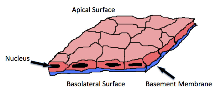
Epithelium:  single or multiple layers of cells that cover body surfaces or line internal cavities and form glands (mirigyek).  They are attached to a basement membrane (alaplemez) of collagen (for strength) and are linked to each other by junctions.  There are no blood vessels in the epithelium, rather nutrient and gas exchange occur by diffusion.  The functions of the epithelium include protection from injury and infection, some absorption, secretion (from glands) and stimulus reception (ingerfelvétel)

Classification
A.  True epithelium (fedőhám):  single cell layer, forms thin linings
i) squamous epithelium (laphám) - flat cells that form thin sheets, permeable to diffusion. eg. line blood capillaries, alveoli (tüdőholyagocskák)


ii) cuboidal epithelium (köbhám) - single layer of cuboid cells, may have microvilli (mikrobolyhok), often involved in absorption and scecretion.  eg. line part of gut, respiratory tract and kidney tubules


iii) columnar epithelium (hengerhám) - single layer of tall cells, their surface is frequently covered with microvilli.  Secretory (goblet) cells are often found between them.  They line the digestive system.


iv) cilliated epithelium (csillóshám) - columnar cells with cilia-covered surfaces.  Line tubes and cavities where materials move.


v) stratified epithelium (többrétegű hám) - multi-layers, forms the epidermis (upper layer of the skin), lines the esophagus and vagina.


B.  Glandular epithelium (mirigyhám)
i) individual glandular cells, eg. goblet cell


ii) multicellular gland - individual goblet cells clustered together.  A gland can be exocrine (külső elválasztású) meaning that it has ducts through which the secretions are released to the surface, for example, sweat glands, or a gland can be endocrine (belső elválasztasú), which means it secretes directly into the blood stream, for example, the thyroid.

2.  Connective and Supportive Tissues (kötő- és támasztószövetek)
These tissues function to bind other tissues and organs together.  They provide protection and support and fill in spaces.  Some of these tissues carry out special roles, such as the storage of fat in fat cells, or those that form blood cells.
Connective tissue is generally formed of networks of various cell types, which often include
fibroblasts

elastin fibers

collagen fibers

macrophages


The most common kinds of connective tissues are:
connective tissue proper
skeletal tissue (cartilage and bone)
blood

a) Connective Tissue Proper (tulajdonképpeni kötőszövet)
- fibroblasts are the predominant cell type.  They produce (secrete) the ground substance and proteins for fibres

- organization:
i) Loose connective tissue (laza rostos kötőszövet)
-thin fibers, not too many fibers, quite a few cells
-found beneath skin, around organs (forms sheets around/between them)

Loose (Areolar) Connective Tissue
This is an example of loose connective tissue. The elastic fibers (EF) are arranged in a random fashion and help the tissue respond to distention. The nucleus of the inactive fibroblast (IFN) is long and flat.
Source: 
http://bcrc.bio.umass.edu/histology/?q=node/180

Loose (Areolar) Connective Tissue
Areolar Connective Tissue: MC=Mast Cell, RF=Reticular Fiber, CF=Collagen Fiber
Source:
http://bcrc.bio.umass.edu/histology/?q=node/343


ii) Adipose Tissue (zsírszövet)
- found in groups surrounded by loose connective tissue
- stores fat (energy reserves)
- also pads some organs (mechanical protection) and insulates from heat loss (thermoregulation)
Source: http://www.deltagen.com/target/histologyatlas/atlas_files/musculoskeletal/adipose_tissue_white_40x.htm


iii)  Dense (fibrous) connective tissue (tömött rostos kötőszövet)
- many fibers, especially collagen.  Fibers are often tightly packed.
-  provides connections between tissues where tension is exerted in a specific direction.  
- examples are:  tendons (inak) and ligaments (izületi szalagok)
- resist tearing
Dense Regular Connective Tissue- Human Tendon
Dense regular connective tissue: human tendon (BV=blood vessel)
Source:  http://bcrc.bio.umass.edu/histology/?q=node/1092

b) Supporting (Skeletal) Tissue (Támasztószövet)
i) Cartilage (porc)
- is a chondrin matrix (jelly-like) embeded with cartilage cells (chondroblasts and chondrocytes), collagen fibers and elastin fibers
hyaline cartilage
Hyaline cartilage (eg. nose, trachea)
Source:  http://legacy.owensboro.kctcs.edu/gcaplan/anat/histology/api%20histo%20connective.htm
elastic cartilage
Elastic cartilage (eg. ears, epiglottis)
Source: http://legacy.owensboro.kctcs.edu/gcaplan/anat/histology/api%20histo%20connective.htm 
ii) Bone (csont)
-  harder than cartilage
- provides protection and a rigid framework
- stores mineral salts
- marrow produces red and white blood cells
- the matrix is imbedded with bone cells (osteoblasts and osteocytes) and collagen
- minerals (CaPO4, CaCO3) deposit around fibers to make bones hard
- cells and fibers provide elasticity
- 2 types of bone:  compact bone and spongy bone
compact bone
-cells are arranged in concentric circles (lamellae) around nerve and blood vessel channels (Haversian canals)
-found in long, shaft bones
The three dimensional structure of compact bone
http://tle.westone.wa.gov.au/content/file/969144ed-0d3b-fa04-2e88-8b23de2a630c/1/human_bio_science_3b.zip/content/003_musculo_skeletal_support/page_05.htm

A labelled micrograph of the internal structure of compact bone showing the osteon, central canal, transverse canal, osteocyte, lamella
http://tle.westone.wa.gov.au/content/file/969144ed-0d3b-fa04-2e88-8b23de2a630c/1/human_bio_science_3b.zip/content/003_musculo_skeletal_support/page_05.htm

spongy bone
- irregular spacing of bone cells
- red marrow fills the spaces (and produces blood cells)
- found at ends of long bones
Trabaculae
SEM of the trabeculae of spongy bone.
x40
Source:  http://www.gla.ac.uk/t4/~fbls/files/fab/tutorial/generic/bonet.html
Picture
Spongy bone 100x
Source:  http://employee.lsc.edu/faculty/BrianBich/Picture%20Library/Forms/DispForm.aspx?ID=578

Compact and spongy bone in the head of the femur
End of femur bone
Source:  http://www.gla.ac.uk/t4/~fbls/files/fab/tutorial/generic/bonet.html
c) Blood
- cells in watery matrix (blood plasma), so they can move.  Plasma also transports nutrients, wastes, hormones, enzymes, dissolved gases, etc
- no fibers
- variety of cell types: 

i) Red blood cells (erythrocytes)
- biconcave disc, has lost nucleus
- contains haemoglobin (Fe gives cell red colour, haemoglobin carries O2)
http://singularityhub.com/2008/08/22/is-an-unlimited-supply-of-blood-and-no-more-need-for-blood-donors-around-the-corner/


ii) White blood cells (leukocytes)
- larger, nucleated cells
- there are fewer in the blood than red blood cells
- great diversity of types:  granulocytes, lymphocytes, etc (more later)
- help in fighting infections
White Blood Cell Photomicrogragh
Source:  http://biology.about.com/od/cellbiology/ss/white-blood-cell.htm

iii.  Platelets (vérlemezkék)
- tiny
-very important in blood clotting
Source:  http://hematologyoutlines.com/atlas_topics/165.html?topic=Agranular%20Platelets*&cb=inline_content_10

3.  Muscle Tissue
a) Skeletal Muscle
-attaches to skeleton (via tendons)
- voluntary movement
- powerful, rapid contractions, but tires quickly
- long, cylindrical cells with striations (stripes), therefore also called striated muscle
- cells are bundled together and enclosed by connective tissue to form a muscle (eg. bicep)

Organization of skeletal muscle
Source:  http://quizlet.com/3238205/muscle-tissue-flash-cards/


Source:  http://faculty.sdmiramar.edu/faculty/sdccd/kpetti/bio160/documents%20biol160.htm

Source:  http://stevegallik.org/sites/histologyolm.stevegallik.org/htmlpages/HOLM_Chapter07_Page04.html
b)  Smooth Muscle
- on walls of internal organs, such as blood vessels, bladder, intestinal wall.
- involuntary muscle, contracts slowly, fatigues slowly, contractions are weak, but long-lasting
- it contains myofibrils, but the organization is different, so no striations are visible.

Source:  http://www.mhhe.com/biosci/ap/histology_mh/nonstria.html

c)  Cardiac muscle (heart)
- striated, but fibers branch at ends and are connected to each other by junctions so signals pass rapidly between cells and they all contract together.
-  rapid, powerful contractions
- rhythmic and involuntary
- do not fatigue.

Source:  http://www.kumc.edu/instruction/medicine/anatomy/histoweb/muscular/muscle14.htm

4.  Nerve Tissue
- makes up the nervous system (brain, spinal cord, ganglia and nerves)

i) Supporting cells
- support, protect and provide nutrients to the neurones
- glial cells in brain and spinal cord: astrocytes (transport of gases, nutrients and wastes to and from 1the blood), oligodendrocytes (form myelin), microglia (role in phagocytosis)

Astrocytes
Source:  http://astrocyte.info/
Source:http://blustein.tripod.com/Oligodendrocytes/oligodendrocytes.htm

Source:  http://missinglink.ucsf.edu/lm/introductionneuropathology/Response%20_to_Injury/Microglia.htm


Source:  http://keck.bioimaging.wisc.edu/lecture-series-2006-2007.html

- Schwann cells are found in the peripheral nervous system where they form the myelin sheath
Source:  http://faculty.southwest.tn.edu/rburkett/A&P1_nervous_system_lab.htm
Source:  http://neurolex.org/wiki/Category:Schwann_Cell

ii) Neurons
- sense stimuli and control reactions to them
- send nerve impulses to organs to make them react
https://www.boundless.com/psychology/the-brain-and-behavior/neurons/introducing-the-neuron/

- impulses flow through a neuron from the dendrite to the axon

brain-neuron-types1
Source:  http://learnzoology.wordpress.com/tag/neuron-tissue/
- Types of neurons are based on their processes
- Bipolar neurons have 1 dendrite and 1 axon
- Unipolar neurons only have on process, typically found in the dorsal root ganglia of the spinal cord (more on that later)
- Multipolar neurons have multiple dendrites and one axon.  This are the most common type of neuron
- Pyramidal cell is a neuron found in the brain with one axon and multiple dendrites.  Its name comes from the triangular shape of the cell body

- 3 kinds:  motor neurons (mozgató), sensory neurons (erző) and interneurons (társító)

types-of-neurons
Source:  http://learnzoology.wordpress.com/tag/neuron-tissue/
- Motor neurons control effector organs, like muscles and glands
- Sensory neurons receive the sensory information from the environment.
- Interneurons connect the sensory and motor neurons to each other.

Neurons are bundled together to form nerves
Nerve fibres may be motor, sensory or mixed, depending on what kind of neurons they contain.