General Information
• Humans
have closed double circuit circulation:
– Closed
means that the blood runs inside of vessels
– Double
refers to the 2 circuits (or loops) that the vessels form
•
PULMONARY CIRCUIT
•
SYSTEMIC CIRCUIT
• The
HEART is the muscular organ that pumps blood
through the vessels
Two
Types of Circulation:
- Pulmonary: pulmonary artery carries deoxygenated blood
to the lungs to release CO2 and pulmonary vein carries oxygenated
blood back to the heart
- Systemic: arteries bring oxygenated blood
to body cells and veins return deoxygenated blood back to heart
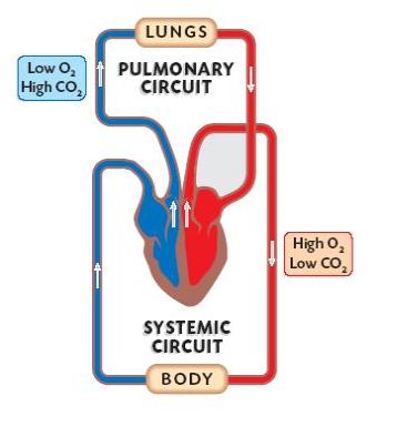 |
| Simple diagram of double circuit circulation |
The Heart (try this interesting link)
 |
- Two
Atria (atrium): upper
two chambers that receive blood. The right atrium receives deoxygenated blood coming from the body (through the vena cava). The left atrium receives oxgenated blood coming from the lungs (through the pulmonary vein).
- Two Ventricles: lower two chambers that pump blood. The right ventricle pumps blood to the lungs (through the pulmonary artery), while the left pumps blood to the body (through the aorta).
 |
 |
The left ventricle is much more muscular than the right ventricle.
 |
| Difference in thickness of the muscular wall of the left and right ventricles |
The right pumps blood to the lungs, while the
left to the entire body. If the
right were as strong as the left, the capillaries of the lungs would explode
when the heart would beat due to the pressure and force behind each pump. The left has to be strong in order to get
blood to all parts of the body efficiently.
Think about the carnival game where you hit the hammer on the lever to
try and get the ball to fly up to the top of the pole and ring the bell. Only the strongest people can do that… your
left ventricle is the “strong man!”
Cardiac cycle is what occurs from 1 heartbeat to the next. It has 2 periods: contraction (systole) and relaxation (diastole). It begins with a rest period when both the atria and the ventricles are relaxed. The atria fill at this point. When the atria contract, but the ventricles remain relaxed, then blood flows into the ventricles. Then the atria relax, but the ventricles contract, pumping blood into the aorta and the pulmonary artery. Then the cycle begins again. The sounds that are heard through the stethoscope are the heart valves closing.
 |
| Cardiac cycle |
 |
Blood
Vessels
Blood vessels carry blood
to every cell in the body
Arteries (osztóerek, verőerek) : carry blood AWAY FROM heart to body.
In the systemic circuit this is oxygen-rich blood, but in the pulmonary circuit this is oxygen-poor blood.
Arteries divide into smaller arteries and arterioles.
Their walls are thick and elastic, so that they can expand and withstand high pressure.
The pulse is the expansion of the artery walls that can be felt easily at the wrists or at the carotid arteries in the neck.
Blood pressure measures 2 values, the systolic pressure (when the left ventricle contracts) which is usually about 120mmHg, and the diastolic pressure (when the left ventricle relaxes), which is usually 80mmHg. The measurement unit is mm of mercury, because the traditional measuring instrument was a sphingomomanometer, which had an arm cuff attached to a tube filled with mercury. Today most are digital.


Blood pressure can only be measured in the arteries. Pressure is very low in the capillaries and veins.
Veins (gyütőerek): carry blood from capillaries BACK TO heart.
In the systemic circuit, this is oxygen-poor blood, but in the pulmonary circuit this is oxygen-rich blood
The smallest veins are called venules and they merge to form veins.
These vessels have a large diamenter, thin walls and are not very elastic
There is very low pressure in the veins, therefore to move blood back to the heart help is required:
a) suction force from the relaxation of the heart (this only works close to the heart),
b) movement of skeletal muscles increases pressure and moves the blood,
c) pocket valves in the veins (especially where blood is moving against gravity) help to keep the blood from running in the "wrong" direction.
Capillaries: microscopic blood vessels connect arteries (arterioles) to veins (venules)
Walls are only ONE CELL THICK!! Specialized for gas and nutrient exchange
Nutrients and oxygen diffuse into body cell
Waste and carbon dioxide diffuse out of body cells
Diameter is so small that red blood cells must travel in single file. Flow is very SLOW.
Plasma flows out into spaces between the cells forming tissue fluid (szövetnedv), which is collected and returned to blood circulation at the venous ends of capillaries or via the lymphatic vascular system (nyirokkeringési rendszer)
Arteries (osztóerek, verőerek) : carry blood AWAY FROM heart to body.
In the systemic circuit this is oxygen-rich blood, but in the pulmonary circuit this is oxygen-poor blood.
Arteries divide into smaller arteries and arterioles.
Their walls are thick and elastic, so that they can expand and withstand high pressure.
 |
Blood pressure measures 2 values, the systolic pressure (when the left ventricle contracts) which is usually about 120mmHg, and the diastolic pressure (when the left ventricle relaxes), which is usually 80mmHg. The measurement unit is mm of mercury, because the traditional measuring instrument was a sphingomomanometer, which had an arm cuff attached to a tube filled with mercury. Today most are digital.
Blood pressure can only be measured in the arteries. Pressure is very low in the capillaries and veins.
 |
| Source: http://classes.midlandstech.edu/carterp/Courses/bio211/chap19/chap19.html |
Veins (gyütőerek): carry blood from capillaries BACK TO heart.
In the systemic circuit, this is oxygen-poor blood, but in the pulmonary circuit this is oxygen-rich blood
The smallest veins are called venules and they merge to form veins.
These vessels have a large diamenter, thin walls and are not very elastic
 |
a) suction force from the relaxation of the heart (this only works close to the heart),
b) movement of skeletal muscles increases pressure and moves the blood,
c) pocket valves in the veins (especially where blood is moving against gravity) help to keep the blood from running in the "wrong" direction.
 |
| Source: http://antranik.org/blood-vessels/ |
Capillaries: microscopic blood vessels connect arteries (arterioles) to veins (venules)
Walls are only ONE CELL THICK!! Specialized for gas and nutrient exchange
Nutrients and oxygen diffuse into body cell
Waste and carbon dioxide diffuse out of body cells
Diameter is so small that red blood cells must travel in single file. Flow is very SLOW.
 |
| Source: http://antranik.org/blood-vessels/ |

Source: http://classes.midlandstech.edu/carterp/Courses/bio211/chap19/chap19.html
At the arteriole end of the capillaries, there are sphincters (ring-like muscles), which open and close to control blood flow to different tissues.
| ||

 |
| Human Circulation |
The above diagram indicates the most important blood vessels . Note the hepatic portal vein which connects the capillaries of the digestive system with those of the liver. The liver filters out toxins and regulates glucose levels. Wastes are filtered out by the kidneys.
Blood
(approx. 5L)
 |
| Contents of blood |
- nutrients (glucose, amino acids, glyercol, fatty acids),
- ions (Na+, K+, Ca2+, Cl-, HCO3-),
- oxygen to cells and
- wastes away (carbon dioxide, carbamide, uric acid). I
- lipids,
- vitamins,
- hormones
- Plasma proteins which include albumin which helps keep the water concentration in blood at that of tissues, globulins which are important in lipid and fat-soluble vitamin transport, and fibrinogen which helps form blood clots.
Their shape is the result of "losing" the nucleus when the rbc matures. Without a nucleus they only live 120 days (broken down in liver and spleen - haem forms bile pigments, globin is recycled)
 |
At high oxygen concentrations (in the lungs), oxyhaemoglobin forms, but at low oxygen concentrations it dissociates to haemoglobin and oxygen (at tissues). Haemoglobin can also bind to CO2 , but to a lesser extent. On the other hand, haemoglobin has a very high affinity for CO (carbon monoxide) and will bind to it in place of oxygen, which is what causes carbon monoxide poisoning.
High level material: CO2 from tissues diffuses into the blood plasma and then into the red blood cells. Inside the cell, the enyzme carbonic anhydrase causes most of the CO2 to react with water to form carbonic acid (H2CO3) which dissociates to form hydrogen ions and hydrogen carbonate (bicarbonate). This is a reversible reaction in a dynamic equilibrium.
H2CO3  H+ + HCO3-
The hydrogen carbonate ions diffuse from the cytoplasm of the red blood cells to the blood plasma. This is balanced by the diffusion of chloride ions in the opposite direction, to maintain a balance of charges on both sides. This is called the "chloride shift".
This dissociation of carbonic acid decreases blood pH (increases acidity). Hydrogen ions react with oxyhaemoglobin to release bound oxygen and reduce the acidity of the blood. This buffering mechanism allows large quantities of carbonic acid to be carried in the blood without significantly changing blood pH.
Hb.4O2 + H+ HHb+ + 4O2
(Hb.4O2 is sometimes written HbO8.)
 |
| Source: http://www.rsc.org/Education/Teachers/Resources/cfb/transport.htm |
White Blood Cells: main function is in defense
There are a variety of types, all large, with irregular shapes
On average, they survive 5-9 days
They are produced in the red bone marrow
They can move out of blood into tissues spaces to reach sites of injury or infection
Types:
- Granulocytes: look granular, include neutrophils, eosinophils and basophils, function in general protection, are phagocytes, so they engulf the "invaders"
- Monocytes: mature into macrophages, they are the most efficient phagocytes, funcion in general defense
- Lymphocytes: specialized to destroy specific foreign particles/viruses/bacteria only after identifying them,
 |
 |
 |
Clotting is referred to as a cascade indicating that if one step is missing the clot doesn't form. It includes many different molecules (referred to as factors) and requires calcium and vitamin K to occur as well. Below is a diagram that shows how complex it is (don't worry about the details, it is just so you understand that it is complex).
 |
Remember blood clotting is an example of positive feedback.
 |
Cardiovascular disease is one of the most common causes of death in Hungary (and in the developed world!). It includes a variety of conditions, which may or may not occur together in one patient, but certainly affect one the other.
1. Arteriosclerosis: occurs when arteries become thick and stiff. This process gradually decreases blood flow to tissues and organs. There are different types that occur depending on the cause of the thickening. Atherosclerosis is the caused by the build up of plaques on the artery walls. The plaques are formed of cholesterol, fatty substances, cellular waste products, calcium and fibrin.
 |
| Source: http://www.taseerlabs.com/MensHealth/7.Blood%20vessel%20abnormalities.html |
 |
3. Heart attack (myocardial infarction): occurs when the heart muscle doesn't get enough oxygen. The heart receives its blood supply through the coronary arteries.
 |
If the coronary arteries have become narrowed due to the build up of plaques along the walls of the arteries, then less oxygenated blood will reach the heart. A clot in a coronary artery can also block the flow of blood to the heart. In a heart attack the cells in the area of the heart that didn't receive enough oxygen will die. After, the damaged tissue will heal, but scar tissue that cannot contract will remain, weakening the heart. Treatment after a heart attack may include lifestyle changes (eg. quit smoking), medication (to help prevent plaque build up, lower blood pressure, etc). If blockage in the coronary arteries requires it, the patient may undergo some surgery, eg. angioplasty or stenting or in more severe cases, bypass surgery.
4. Stroke: poor blood flow to the brain results in cell death. There are two kinds of strokes: ischemic (more common, but less deadly), when blood is blocked from reaching the brain, and hemorrhagic, due to bleeding in the brain. Effects of a stroke depend on what part of the brain was affected and the extent of the damage.
 |
No comments:
Post a Comment