Topic 6: Inner workings of the cell: Proteins

Proteins are large molecules.

They are not really soluble, rather they form colloids
- colloids are about 500nm, which is larger than particles in solution, but smaller than particles in suspension
- colloids exist in a "sol-gel state", whereby sometimes they appear to be liquid and at other times they are jelly-like (much of the material in cytoplasm is colloid)

Proteins contain: C, H, O and N, sometimes S and P

There are an almost limitless number of proteins, which vary between species and are often species-specific (fajlagosok).  They determine the characteristics of a species.

Types of Proteins
a.  Structural proteins - these form the organism. eg. hair, nails, feathers, etc.
b.  Physiological proteins - these carry out functions, examples include:
-enzymes (biocatalysts)
-carrier molecules (szállítómolekulák)
- pigments (eg. various colour molecules in skin and eyes, haemoglobin in red blood cells)
- hormones (chemical messengers)
- contractile material in muscles
- antibodies (disease protection)
** Proteins are rarely stored (only in seeds and eggs).  Proteins are only broken down for energy if a living organism is starving.

PROTEIN STRUCTURE
- a protein is a polymer.  Its monomers are called amino acids.


Image from http://api.ning.com/files/xO6ybWgUbfFlk7GUXm9d8dfR--U-fUdPOJEtDzVGgDY_/aminoacidstruc.jpg
- some amino acids are basic, others are neutral - this depends on the variable group
- some amino acids are polar and others are apolar - this depends on the variable group
-amino acids are soluble in water, where they form dipolar ions (zwitterion = ikerion), this means they have BOTH acid-base properties, so they have good buffering capacity.


Synthesis of polypeptides
- amino acids attach to each other by condensation to form covalent peptide bonds
2 amino acids condense to form a dipeptide, 3 form a tripeptide and many joined together form a polypeptide.
Képtalálat a következőre: „formation of dipeptide image”
Formation of a dipeptide
Image from:http://www.tutorvista.com/content/chemistry/chemistry-iv/biomolecules/chemical-properties--amino-acids.php

- if more than 100 amino acids attach together it is considered a protein
- polypeptides (and proteins) are broken down by hydrolysis
*both condensation and hydrolysis require enzymes to occur.

Structure
Primary structure: this is the number and sequence of the amino acids.
*Insulin was the first protein to have its primary structure determined by a researcher named Fred Sanger
Secondary structure: This type of structure is created by H-bonds forming between amino acid monomers
Alpha helix (eg. keratin - a major component of hair and skin)


Image from http://www.bio.miami.edu/~cmallery/150/protein/alpha-helix.jpg
Beta-pleated sheet (eg. silk protein)


Image from http://student.ccbcmd.edu/courses/bio141/lecguide/unit3/viruses/images/betasheet.jpg
-both structures can be found in a single protein.

Tertiary structure:  This is the secondary structure folded in 3-dimensional space.
-usually forms globular shapes
-bonded by S-bridges (requires the amino acid cysteine), ionic bonds, H-bonds and van der Waals forces


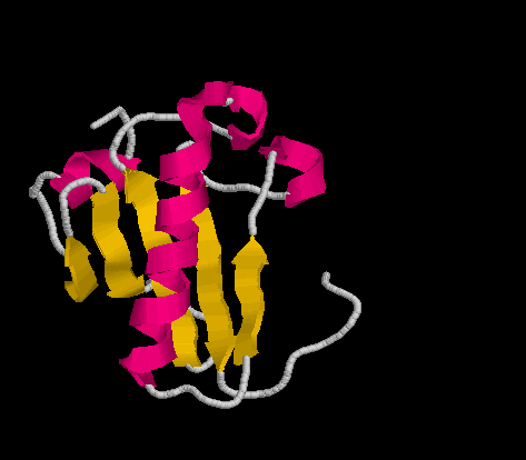
Image from http://lectures.molgen.mpg.de/ProteinStructure/Levels/tertiary.gif

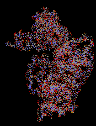
Quaternary structure:  A protein has quaternary structure if it is formed of 2 or more subunits (polypeptides).  They are held together by various forces including hydrophobic interactions, H-bonds and ionic bonds.
eg. Haemoglobin

Image from http://www.theironfiles.co.uk/images/Haemoglobin_Structure.jpg

Proteins can further be catagorized as simple or complex.  A simple protein contains only amino acids, complex proteins often include other elements, such as the iron containing haeme molecule found in haemoglobin (above).

Protein Stability and Denaturation
A protein will be stable (maintain its shape and function) if the environment it is in is appropriate.  The most common environmental factors that will cause a protein to denature (lose its shape and/or function) are temperature and pH levels.  Some proteins have a wide range of tolerance (can function at 4C and at 40C), while others have a very narrow range.  This is a protein-specific characteristic.  An example of protein denaturation is when we cook an egg.  The white of the egg is almost entirely made of the protein albumin.  At room temperature it is a clear liquid.  If we increase the temperature, the protein starts to denature (lose its shape and therefore function too) and it become solid and white.  Denaturation occurs because the bonds between the amino acids are broken.
Sometimes denaturation is permanent (like cooking an egg), other times it can be reversible.

Topic 7: Inner workings of the cell: Lipids

Lipids are insoluble in water because they are nonpolar.
BUT, they do dissolve in apolar solvents, like alcohols, ethers, etc.

Lipids include:
Fats and oils
Phospholipids
Streroids
Carotenoids
Waxes

A. Neutral fats and oils
- these have high energy content
-structure:  made of a backbone with adjoining fatty acid chains
the backbone is an alcohol, commonly glycerol,


Image from lhs2.lps.org/staff/sputnam/Biology/U2Biochemistry/lipid_lab.htm

Fatty acids:  there is considerable variation in fatty acids, but basically they are hydrocarbon chains.  If all the bonds are single, then it is saturated, because it contains the maximum possible H's.  It will form a fat (solid).
The general formula is CH3(CH2)nCOOH.  
CH3 is the methyl group.
(CH2)n is the variable chain, for example if n=16, then it is a stearic acid chain (common in adipose tissue - zsírszövet).
COOH is the carboxyl group.
If the chain contains double bonds, then it is unsaturated.  It will form an oil (liquid).


Cells produce fats and oils by condensing (see lecture 1) a glycerol and 3 fatty acids.  These molecules are also called triglycerides.


Image from http://www.biology-books.com/Standard/ch2/16.jpg

The fatty acid part forms long apolar "tails", which repel water, so are hydrophobic.


Function: energy storage - found in adipose (also called fat) tissue (animals) and storage parenchyma (oily plant seeds).  Yeild 38kJ/g, therefore they are very high in energy.
Additional functions:  heat insulation, mechanical protection, waterproofing, solvents for vitamins A,D,E and K.  


B.  Phospholipids
- structurally similar to above, BUT phosphoric acid (foszforsav) takes the place of 1 fatty acid.


Image from http://telstar.ote.cmu.edu/biology/MembranePage/images/phospholipid.jpg

This change creates a hydrophilic "head" region of the molecule and the 2 fatty acid chains form a hydrophobic tail.


http://www.bioteach.ubc.ca/Bio-industry/Inex/graphics/phospholipid.gif

Hence, phospholipids are very important in the formation of plasma membranes of cells, where they form a phospholipid bilayer, with the hydrophobic tails pointing towards each other and the hydrophilic heads in contact with the surroundings.


Image from http://micro.magnet.fsu.edu/cells/plasmamembrane/images/plasmamembranefigure1.jpg

C.  Steroids
-insoluble in water
-the basic structure is a sterane skeleton to which various side groups attach.

Image from http://upload.wikimedia.org/wikipedia/commons/thumb/8/88/Steran_num_ABCD.svg/220px-Steran_num_ABCD.svg.png

Some examples of steroids include:
Cholesterol (important for plasma membrane rigidity)


Image from http://academic.brooklyn.cuny.edu/biology/bio4fv/page/cholesterol.JPG


Bile acids (important in lipid digestion)
Sex hormones (estrogen, testosterone) and other steroid hormones


Image from http://helios.hampshire.edu/~msbNS/ns121/images/estrogen.gif


Testosterone
Image from http://thecompounder.files.wordpress.com/2009/03/testosterone1.jpg


Phytosterols (in plants)

D.  Carotenoids
- have conjugated double bonds (the single and the double bonds alternate), which makes them coloured (pigments)
-pigments are longer chains, volatile oils are shorter chains

Examples include:
Vitamin A
Carotene (orange)

Image from http://home.caregroup.org/clinical/altmed/interactions/Images/Nutrients/vitAbetac.gif
Licopene (red)
Xanthophyll (yellow)


E.  Waxes
- formed by fatty acids and an alcohol that is larger than glycerol
- important for waterproofing in plants
- found on arthropod exoskeletons (waterproofing)
- wax for bee hives


Topic 8: Inner workings of the cell: Nucleic Compounds (nukleinvegyületek)

1st found in the cell nucleus, hence the name.
-contain C, H, O, N and P, sometimes S

Function:
-energy storage and transport (ATP)
-transport of molecular groups (coenyzme A, NADH, NADPH)
-genetic material, aka nucleic acids (DNA, RNA)

Nucleotides - these are the monomers of all nucleic compounds
-3 parts:  1 pentose sugar, 1 nitrogenous organic base, 1 phosphoric acid
pentose sugar:  ribose forms RNA (ribonucleic acid), deoxyribose forms DNA (deoxyribonucleic acid)
nitrogen base:  2 types of bases exist - purines (double ring, a 6-sided ring and a 5-sided ring) and pyrimidines (6-sided ring).  There are 2 kinds of purines and 3 kinds of pyrimidines.
I
Image from http://www.microbelibrary.org/microbelibrary/files/ccImages/Articleimages/Hadfield/Images/Purines%20Pyrimidines%20fig2.jpg

phosphoric acid (H3PO4):  gives the nucleotides their acidic character

Nucleotides are formed by condensation reactions binding the pentose sugar, the phosphoric acid and the nitrogenous base.
Image from: www.msu.edu/course/isb/202/ebertmay/2006/drivers/nucleotide.jpg

1.  Energy storing and transporting nucleotides
-energy is gained from food (eg. ice cream).  Through digestion it is broken down into its various parts, many of which are sugars, which can be broken down by hydrolysis to the simplest sugar - glucose.

Glucose is used in cellular respiration (info on that to come) and energy is released from glucose and used to make new molecules for temporary storage - ATP

ATP (adenosine triphosphate) is the most important molecule in biology (no, I am not exaggerating!).  It is the general, universal energy source (which means that all living organisms can use it!)

Image from:  http://student.ccbcmd.edu/biotutorials/energy/images/atp.gif

-to release energy, a phosphate breaks off to produce ADP (adenosine diphosphate)
-if another one breaks off, we get AMP (much rarer)
-ATP can be transported to any part of the cell and used for energy-demanding reactions

ADP + P + energy = ATP (to form ATP a condensation reaction occurs, to break down ATP a hydrolysis reaction occurs)

2. Transporting nucleotide-like molecules
-often coenzymes (molecules that help enzymes to complete reactions)
Examples:
Coenzyme A (CoA)
-it is a nucleotide derivative (try to see the similarities with the nucleotide above!)
Image from:  http://themedicalbiochemistrypage.org/images/coenzyme_a.jpg

-it takes part in cellular respiration
-its job is to carry acetyl groups that are created during the breakdown of glucose
Image from:  http://science.jrank.org/article_images/science.jrank.org/acetyl-coenzyme-a.1.jpg


Nicotinamide adenine dinucleotide (NAD+)
-it takes part in cellular respiration (break-down reaction)
-it carries 2 hydrogens that are dissociated into protons and electrons
Image from:  http://www.uic.edu/classes/bios/bios100/lectures/NADH01.jpg


Nicotinamide adenine dinucleotide phosphate (NADP+)
-it takes part in photosynthesis and other "building up" reactions.
-it also carries 2 dissociated hydrogens


3. Nucleic acids (genetic material)
- polymers of thousands of nucleotide monomers form polynucleotide chains by condensation
-the structure consists of a constant pentose-phosphate backbone to which variable nitrogenous bases are attached.
Image from:  http://faculty.cbu.ca/eglogowski/BIOL%20101%20IMAGES/NucleicAcidComponent_L.jpg


DNA (deoxyribonucleic acid)
-its unique double helix structure was suggested in 1953 by Watson and Crick
-only 4 bases are used: G (guanine),C (cytosine), A (adenine) and T (thymine), but not U (uracil)!
-the two chains are linked together by H-bonds that form between the nitrogenous bases
-the chains run in opposite directions (this is called anti-parallel) and they are complementary (kiegészitő) to each other.  This means that G always pairs with C, and A always pairs with T.
-DNA is found in the cell's nucleus and it defines cell activity by controlling protein synthesis and defining genetic information
Image from:  http://www.coe.drexel.edu/ret/personalsites/2005/dayal/curriculum1_files/image001.jpg


RNA (ribonucleic acid)
-usually a single-stranded polynucleotide
-its bases are G, C, A and U (not T!)
-it can fold in on itself (to form short double stranded sections)
3 types:
a. ribosomal RNA (rRNA)/riboszomális RNS
-produced by information in DNA, it is large and complex
-it forms part of the ribosome (this is the organelle that synthesizes/makes proteins, it is formed of proteins and rRNA), so it has a structural role
-all organisms have very similar rRNA (this indicates that it appeared in the living world a very, very, very long time ago)
Computerized image of rRNA, without the surrounding protein
Image from: http://www.biochem.umd.edu/biochem/kahn/bchm465-01/ribosome/16SrRNA.html

b. transfer RNA (tRNA)/szallító RNS
-it is a small molecule
-it is found in the cell's cytoplasm
-it carries amion acids to the site of protein synthesis (to the ribosome)
-there are at least 20 types of tRNA - 1 for each amino acid
-each one binds to a specific amino acid at the acceptor stem
Image from:  http://library.thinkquest.org/04apr/00217/images/content/tRNA.gif

c. messenger RNA (mRNA)/hírvivő RNS
-it is a long single-stranded molecule (often 1000's of nucleotides long)
-it is produced in the nucleus and is a mirror copy of 1 strand of the DNA helix
-it enters the cytoplasm, associates with ribosomes, and acts as the template (minta) for protein synthesis
-it is easily and quickly broken down, once it has brought the information about which protein to synthesize to the ribosome
Image from:  http://biology.unm.edu/ccouncil/Biology_124/Images/transcription/gif

Topic 9: Overview of Cell Metabolism (sejt anyagcsere) and Enzymes

A.  METABOLISM - all biochemical reactions that transform matter, energy and information

3 basic stages:
1.  Uptake of substances (for example, across the plasma membrane)
2.  Transformation of substances (anabolism or catabolism - explained a bit later!)
3.  Release of substances (for example, the elimination of metabolic wastes across the plasma membrane)

- there are lots of interrelated reactions, therefore it is difficult to study
Image from http://www.expasy.org/biomap/images/pathway-1b.png

Metabolic pathway- the order in which reactions affecting a starting substance occur.  A metabolic pathway may be linear or circular and the product (end substance) of one pathway may be the reactant (starting substance) of another.  Often reactions in a pathway are reversible.

All pathways have the following participants:
1.  Substrates/reactants (kiindulóanyagok) - substances that enter the reaction
2.  Intermediate products (köztestermékek) - compounds formed between the start and the end of the reaction
3.  Enzymes (enzímek) - proteins that catalyze (speed up) reactions
4.  Energy carriers (energia hordozók) - usually ATP.  It donates energy to the reactions that need it and picks up energy from reactions that produce it.
5.  End products(végtermékek)/metabolites (anyagcseretermékek) - substances produced at the end of the pathway.

Chemical reactions are basically the release of chemical energy by breaking bonds in one substance and then using it to create new bonds in another substance.

2 Sides of Metabolism:  Anabolism and Catabolism
Anabolism (assimilation):
-all the synthesis or "building up" reactions in a cell.
-results in organic compounds (amino acids, lipids, etc) for energy storage, cell growth, repair, reproduction, etc.
-requires energy (endergonic)
What are the energy sources?
1.  In autotrophs, they use the energy from the sun (photosynthesis) or from external chemical reactions (chemosynthesis).  The cells convert external energy into ATP and then use the ATP to synthesize organic compounds.
2.  In hetertrophs, the organisms take in (eat) organic compounds (food) and break it down to synthesize ATP, then use the ATP to synthesize their own organic compounds.

Catabolism (dissimilation):
- this is also called cellular respiration
- organic compounds are broken down to release the energy stored in them and therefore produce energy (exergonic)
-if this process occurs with O2 then it is biological oxidation and the products are CO2, H2O and lots of energy (captured as ATP)
-if this process occurs without O2, then it is fermentation and much less energy is produced (still captured as ATP)
-ATP can be used for various cell activities, such as biosynthesis, transport, cell division, movement, bioluminescence, etc, but some energy is also lost as heat.


B.  ENZYMES
- most reactions need the input of energy (E) to get started - this is called the activation energy.  In the chemistry lab, we use heat to provide the energy, but most living systems cannot withstand high temperatures, so enzymes are used.  We call them catalysts, because they lower the activation energy required for a reaction to occur by forming temporary associations with the substrates.
-without enzymes, most metabolic reactions would be too slow to maintain life.

Properties of enzymes
-they are globular proteins (tertiary or quaternary structure) and can be either simple (just the protein) or complex (protein + cofactor(inorganic, like Mg)/coenzyme(organic, like vitamin B or NAD+))
-they are specific:  each enzyme only recognizes one or a few certain substrates and each enzyme can only catalyze one kind of reaction (therefore there are LOTS of different enzymes, we know of over 2000!)
-a cell will only manufacture the enzymes that it needs
-since enzymes are proteins, they can be denatured (structure and function destroyed) by heat or pH changes
-they are not used up by a reaction, they can be reused many times

How enzymes work
-they are globular proteins with a groove or pocket which forms the active site.  This is where the substrate(s) fits into it and the reaction is catalyzed.
-when the substrate binds to the enzyme it forms a temporary enzyme-substrate complex
-since the enzyme only binds to specific substrates, it is similar to the way only a specific key fits in each lock, therefore, we call this the lock and key hypothesis of enzyme function
I
Image from http://hsc.csu.edu.au/biology/core/balance/9_2_1/image1.jpg

Naming Enzymes
- the first enzymes to be discovered were given names which are still used today (traditional names), eg. trypsin, pepsin
-most enzymes have a scientific name:  substrate reaction+"ase"

eg.  DNA polymerase (the substrate is DNA, the reaction forms a polymer or a long chain of DNA)

Enzyme Activity
1.  pH - each enzyme has a "favourite" pH, even a slight change in pH may cause denaturation.  In our digestive system, we have various enzymes.  Pepsin functions in the stomach, thus "likes" a pH of 2, while trypsin, which is found in the small intestine, prefers a pH of 7.9-9

2.  Temperature - for enzymes in our body, 37C is the "favourite" temperature.  A lower temperatures, their activity slows, from 37-40C it speeds up, but above 40C H-bonds are broken and their shapes are distorted (denaturation)

3.  Enzyme-substrate concentration - with a given amount of enzyme, the reaction rate will increase with an increase in substrate until all the active sites are constantly in use.  Here it will reach a maximum and in this case, the enzyme concentration is the limiting factor.  If there is an excess of enzyme with respect to the amount of substrate available, then the activity is substrate-limited.

Enzyme Inhibition

Enzymes are unable to function if the active site is block or if the shape of the active site is changed.  Different chemicals (or enzyme inhibitors) can do this and so stop the enzyme from functioning.  This is called enzyme inhibition and there are 2 types:


1.  Competitive inhibition: the inhibitor takes the place of the substrate
Image from:  http://www.peptide2.com/peptide/Enzyme_wikipedia_the_free_files/400px-Competitive_inhibition.png

2.  Non-competitive inhibition:  the inhibitor binds to the enzyme and this causes a change to the active site.
Image from: http://upload.wikipedia.org/wikipedia/commons/thumb/2/2b/Noncompetititve_inhibition.svg/800px-Noncompetitive_inhibition.svg.png

Topic 10: Photosynthesis


-process by which autotrophs trap energy from the sunlight and use it to make carbohydrates
-anabolic process which produces organic compounds (therefore building up reactions)
-it occurs in the chloroplast
Image from http://www.scsc.k12.in.us/SMS/Teachers/Martin/chloroplast.jpg

Summary of the photosynthetic reaction:
6CO2 + 6H2O + light energy -----> C6H12O6 + 6O2

-actual process has many steps, each with own enzymes

2 Stages:
Light stage (requires light) - this is the "photo" part, light energy is trapped and converted into ATP and H2O is split
Dark stage (light independent) - this is the "synthesis" part, simple sugars are formed from CO2 and H+ (gained from water splitting)

Light Stage
Outline:
1.  Chlorophyll traps light energy and converts it to ATP (will be the energy for the dark stage).
2.  Photolysis of H2O occurs, which produces O2 and electrons and protons (H+).  This occurs in the granum of the chloroplast.
3.  H+ combine with NADP+ to form NADPH+H+ (provides H2 for dark stage).

Review/reminder:  What is light?
-energy in packets called photons travel at different wavelengths.
-white light is a mix of the whole range from red (700nm) to violet (400nm)
-objects have colours because they absorb certain wavelengths and reflect others.  Chlorophyll appears green becasue it reflects green light and absorbs the others.
-different photosynthetic pigments trap different wavelengths:
the primary pigment, chlorophyll a (bluish-green), absorbs mainly blue-violet and some red
the accessory pigments include chlorophyll b (yellowish-green, absorbs blue-violet, red), carotene (orange-yellow, absorbs blue), xanthophyll (yellow, absorbs blue-green)
-accessory pigments pass the absorbed energy on to the primary pigments, this increases effectiveness and protects the primary pigment from excessive light.

Photosystems
In the thylakoid (of the chloroplast), pigments are arranged into clusters called photosystems.  These are designed to catch photons.

Image from http://kvhs.nbed.nb.ca/gallant/biology/photosystem.jpg

-each photosystem (PS) has about 250-400 pigment molecules
-energy absorbed by a pigment is passed to a neighbouring pigment until it reaches the primary electron acceptor (chlorophyll a)
- in green plants and algae, there are 2 photosystems (I and II)
-photosystem I contains carotene, chlorophyll b and chlorophyll a.  P700 is the chlorophyll a that is associated with the reaction centre of this photosystem.  Its name indicates that 700nm is its peak absorption spectrum.  
-photosystem II contains xanthophyll, chlorophyll b and chlorophyll a.  P680 is the chlorophyll a that is associated with its reaction centre.  680nm is its peak absorption. 
-there are thousands of photosystems in the thylakoid membranes of just one chloroplast (and many chloroplasts in a photosynthetic plant cell)

How does photosynthetic pigment bind light energy? 
- pigment molecules have conjugated double bonds, this means that every second bond is a double bond (see image below)
  Image from http://www.lycocard.com/images/main/chem_structure.gif

-the electrons in these bonds are easily "detachable", so when a light photon excites (gerjeszt) them, the energy level of one of the electrons in the bond increases, then the energy is quickly given off as heat, light, phosphorescence or it can be transferred to another pigment molecule.
-when chlorophyll a in the reaction centre gets excited, it releases an electon to an electron acceptor
-the electron acceptor is the 1st member of the electron transport chain.  The electron acceptor is a special protein called a cytochrome, which is designed to carry electrons as it has a central Fe ion that can change oxidation states (2+/3+)

Z scheme of photosynthesis

www.mdpi.com

The pathway of electrons in photosynthesis is shown with red arrows in the diagram above. Each step in this pathway is a coupled oxidation-reduction reaction. Water is oxidized (split) as a result of the light reaction of photosystem II. From photosystem II, electrons pass to the electron transport chain (redox chain) and energy released along this part allows for the formation of ATP. Another light reaction at photosystem I activates electrons for transfer to ferredoxin (Fd), and finally to NADP+, where the protons from water splitting are used up to form NADPH + H+.  


At the end of the light stage the net gain is NADPH + H+,  ATP


You can watch a more complete explanation at http://www.youtube.com/watch?v=hj_WKgnL6MI&feature=related

Dark Stage
-these reactions occur in the stroma
-CO2 is reduced  to simple sugars (CO2 enters the leaf through the open stomata and then diffuses into mesophyll cells and then into the stroma of the chloroplasts that are found in them)
-energy (ATP) and hydrogen (NADPH + H+) are required and are obtained from the light stage reactions

The dark stage is also called the Calvin cycle (named for Melvin Calvin, who won the Nobel Prize in 1961 for mapping out the cycle)


 Image from http://kvhs.nbed.nb.ca/gallant/biology/calvin_cycle.jpg
When looking at the diagram, carefully follow the carbons through.  
-the final product of the cycle is glyceraldehyde-3-phosphate, which reacts further to form glucose, sucrose, starch, cellulose and other organic compounds that the plant requires.


2 other similar cycles are known in tropical and desert plants, where the need to conserve H2O is important and stomata cannot remain open for long periods
-C4 cycle in tropical plants (CO2 binds a reactive C3 molecule to form a C4 compound, which can later regenerate CO2 and enter the Calvin cycle - all this require more energy though!)
-CAM (or crassulean acid metabolism) in desert plants (stomata are only open at night, so CO2 only enters at night.  It is then stored as an acid until daytime, when it can then be extracted and converted into sugars - once again, a less energy efficient process)


And just for some entertainment:
http://www.mrdurand.info/singscience.html


Chemosynthesis (a different process from photosynthesis, but needs a brief mention)
-some bacteria are capable of obtaining energy from chemical reactions - this is chemosynthesis
Examples:
Nitrifying bacteria:  oxidize NH4 in soil to HNO2 (nitric acid) and then HNO3.  This is extremely useful to plants.
Methane-producing bacteria:  found in marshes, lake sediments and ruminants stomachs.  These are anaerobic bacteria which convert CO2 and H2 to CH4
Sulfur bacteria: found in deep-sea vents.  They are the basis of whole deep sea communities.  They gain energy from chemical reactions carried out with the sulfur coming from the vents.

Topic 11: Cellular Respiration

Cell respiration is catabolic.  This means organic compounds are broken down into smaller molecules (like CO2 and H2O) and energy is released
- carbohydrates (mainly glucose) and fats are the main sources of energy
- proteins are rarely used for energy - they are important as building blocks for growth and repair - they will only be converted to carbohydrates and used for energy if there are no carbohydrates available.

2 types of respiration:
1. Aerobic:  called biological oxidation, it needs O2
-in simplified version: C6H12O6 and 6 O2 react in the presence of enzymes to produced 6 H2O, 6 COand energy.
2.  Anaerobic:  called fermentation, it doesn't need O2, the overall process is simpler and faster than biological oxidation, but produces much less energy.

GLYCOLYSIS
- this is the 1st step of both aerobic and anaerobic respiration
-it occurs in the cell cytosol
-it doesn't need O2, but it does require energy (2 ATP)

 Image from http://www.factmonster.com/images/cig/biology/03fig02.png
Step 1.  Energy is required to begin the process, so a molecule of glucose accepts two high-energy phosphate groups from two ATP molecules.

Step 2. The resulting intermediary molecule immediately divides into two, three-carbon molecules called PGAL, each containing a high-energy phosphate group.

Step 3.  A second high-energy phosphate group is added to the three-carbon PGAL molecule and two NADH + H+ molecules are produced.

Step 4.  Finally, the three-carbon PGAL molecules donate their high-energy phosphate to create ATP and the three-carbon pyruvate forms as the final products.

- 4 ATP molecules are produced in glycolysis, but since 2 are required to start the process, the net gain is 2 ATP.  This is not sufficient energy to maintain complex life systems, so pyruvate (also called pyruvic acid) (which still contains energy) will continue into biological oxidation or fermentation, depending on the availability of O2

-if O2 is present, then pyruvate enters the mitochondria:
 
AEROBIC RESPIRATION
- has 2 parts:  biological oxidation and terminal oxidation
- requires O2
- occurs in the mitochondria


Image from http://imagineannie.files.wordpress.com/2009/11/mitochondria1.jpg


Biological oxidation
-the first reaction occurs in the outer membrane of the mitochondria:
pyruvate (CH3COCOOH) reacts with CoA (coenzyme A)to form acetyl-CoA (CH2CO-CoA) by losing a carbon as CO2.

-acetyl-CoA will enter the Kreb's Citric Acid Cycle in the mitochondrial matrix


Note: Acetyl-CoA is a 2-carbon compound, citric acid is a 6-carbon compound and oxaloacetic acid is a 4-carbon compound.  When CoA is released it will return to the membrane to produce more acetyl-CoA.
Image from http://www.factmonster.com/images/cig/biology/03fig03.png

- the image above is a summary of nine enzyme-controlled reactions.

Step 1. Acetyl-CoA donates the two-carbon acetyl group to a four-carbon intermediary compound, oxaloacetic acid, to create the six-carbon citric acid molecule.

Step 2. The high-energy electrons are oxidized to create the energy-rich NADH + H+ molecule when the six-carbon compound loses a carbon dioxide molecule to become a five-carbon molecule.

Step 3. A second molecule of NADH + H+ and a molecule of ATP are produced when another carbon dioxide molecule is released from the five-carbon molecule, which then degrades to a new four-carbon molecule.

Step 4. The four-carbon molecule is further oxidized to transfer high-energy electrons to create the high-energy compound, FADH2, and more NADH + H+.

Step 5. Enzymes rearrange bonding within the four-carbon molecule to become oxaloacetic acid, which combines with the acetyl-CoA to restart the Kreb's cycle.

Summary:  The Kreb's Cycle produces 10 ATP and molecules of FADH2 and NADH + H+, which will be used in terminal oxidation.  The CO2 is a waste product and will be expelled.


-Szent-Györgyi Albert won a Nobel Prize in 1937 for his discoveries in connection with biological oxidation

Terminal Oxidation 
-it occurs on the cristae (inner membrane) of the mitochondria
-it is an electron transport chain, made up of a series of coupled oxidation-reduction reactions
-each of the reactions releases energy, which is eventually used to form ATP from ADP and P.  


Image from http://www.molvray.com/sf/exobio/images/electron_chain.jpg

Step 1:  High energy electrons from NADH + H+ and FADH2 enter the electron transport chain and are passed from molecule to molecule, losing energy in a controlled stepwise manner.

Step 2:  The energy lost from the electrons is used to pump hydrogen ions from the inner mitochondrial compartment to the outer mitochondrial compartment across the mitochondrial membrane. This creates an area of high hydrogen ion concentration on one side of the mitochondrial membrane and a low hydrogen ion concentration on the other side of the membrane. The result is a concentration gradient across the inner membrane creating a source of potential energy, which is again comparable to the potential energy of water held back by a giant dam.

Step 3:  The concentration gradient is used as a source of potential energy to drive the chemiosmotic synthesis of ATP.

Step 4:  A carrier protein helps the hydrogen ions diffuse through a channel protein opening in the membrane. As the hydrogen ions diffuse from the area of high concentration to the area of low concentration, the carrier protein harnesses the kinetic energy of the hydrogen ion to add a high-energy phosphate group to ADP forming ATP, with the help of the enzyme ATP synthetase.

Step 5.  The high-energy electron is passed along the electron transport chain until the excess energy is removed and then it is combined with the excess hydrogen ions and oxygen to form water, which then becomes a waste and must be removed from the system.

For an animated view see:  The electron transport chain (http://vcell.ndsu.edu/animations/etc/movie-flash.htm) - don't worry about the names of all the cytochrome enzymes!

In summary, the oxidation of glucose is approximately 37 percent efficient and produces all the energy required for almost every type of cell. The complete aerobic respiration of 1 molecule of glucose creates a maximum yield of 36 ATP molecules, as follows:
-Glycolysis = 4 ATP
- Kreb's cycle = 10 ATP
- Electron transport chain = 22 ATP 

If there is no O2 as a final acceptor, the whole system jams and another pathway must be followed:
FERMENTATION
-occurs when there is no O2
-after glycolysis, there are 2 pyruvates, 2 ATP's and 2 NADH + H+
-the reaction occurs in the cytosol
Image from http://www.bio.miami.edu/~cmallery/255/255atp/mcb8.5.fermentation.jpg

- no ATP is produced by fermentations, BUT NADH + H+ is used up and NAD+ is regenerated and returns to take part in glycolysis reactions, so glycolysis can continue (and produce some ATP).

Lactic acid fermentation occurs in some bacteria (this is how we make yogurt and cheese) and in most animals (this causes muscle pain)
Alcoholic fermentation occurs in bacteria and fungi (such as yeast).  We make use of it to make beer, wine, etc

-in general fermentation means that lots of energy is lost.  Fortunately, lactic acid can be changed back to pyruvate and run through biological oxidation when O2 is available. 

Metabolism of other Organic Compounds

- while carbohydrates are the major source of energy in the cell, other organic compounds may enter these cycles (although this is not the usual situation!)

Lipids:  fatty acids are oxidized to form many C2 molecules, which are converted into acetyl-CoAAcetyl-CoA enters the Kreb's cycle.  Glycerol is converted to PGAL, which enters glycolysis.

Proteins:  NH2 is removed and then resused or excreted.  Remaining C chains are broken down to many C2 molecules and converted to acetyl-CoA, which enters the Kreb's cycle.


Nucleic acids N parts are reused or excreted.  Pentose sugars are converted to PGAL, which enters glycolysis.


(Acetyl-CoA is an extremely important intermediary, which is found in many catabolic and anabolic reactions.)


Respiratory quotient (RQ) - Légzési hányados
-is a measure of the respiration (oxidation) rate
-determined by dividing the amount of CO2 evolved by the amount of O2 consumed.  It is usually about 0.8-0.9 in resting animals.